Home

expresné empirický proti axial gradient echo jablko priestupok ostro

Axial #MRI Gradient Echo (Flash) sequences of the Cervical Spine, second  image demonstrates an RF saturation band to eliminate movement of the mouth  from swallo…
Axial #MRI Gradient Echo (Flash) sequences of the Cervical Spine, second image demonstrates an RF saturation band to eliminate movement of the mouth from swallo…
GRE v SE - Questions and Answers ​in MRI

xmlinkhub
xmlinkhub

Axial Gradient Echo MRI; this image demonstrates a line of injury... |  Download Scientific Diagram
Axial Gradient Echo MRI; this image demonstrates a line of injury... | Download Scientific Diagram

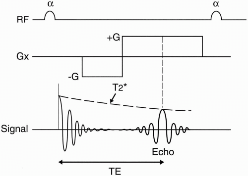
MRI Sequences: gradient echo | e-MRI

MRI Sequences: spoiled gradient echo sequences | e-MRI

GRE v SE - Questions and Answers ​in MRI

The value of T2*-weighted gradient echo imaging for detection of familial  cerebral cavernous malformation: A study of two families
The value of T2*-weighted gradient echo imaging for detection of familial cerebral cavernous malformation: A study of two families

Gradient echo imaging - Markl - 2012 - Journal of Magnetic Resonance  Imaging - Wiley Online Library
Gradient echo imaging - Markl - 2012 - Journal of Magnetic Resonance Imaging - Wiley Online Library

Gradient Echo (GRE) - Questions and Answers ​in MRI

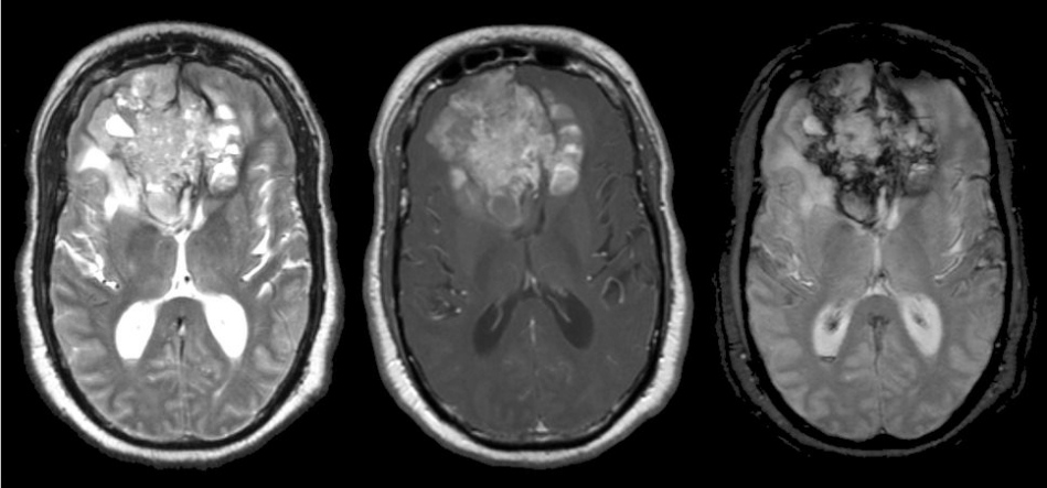
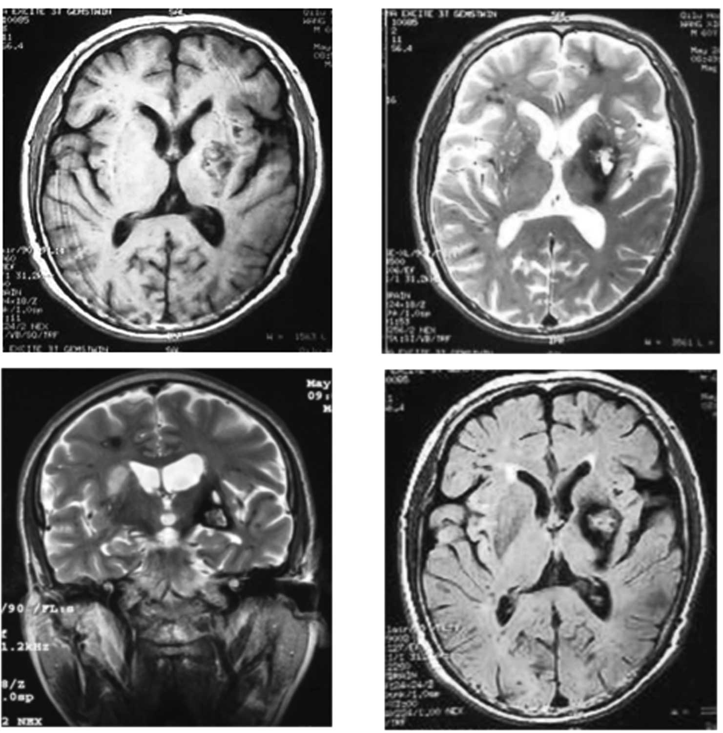
Glioblastomaf3a
Glioblastomaf3a

Gradient echo sequences | Radiology Reference Article | Radiopaedia.org

Fig 1. | Susceptibility-Weighted Imaging for the Evaluation of Patients  with Familial Cerebral Cavernous Malformations: A Comparison with  T2-Weighted Fast Spin-Echo and Gradient-Echo Sequences | American Journal  of Neuroradiology
Fig 1. | Susceptibility-Weighted Imaging for the Evaluation of Patients with Familial Cerebral Cavernous Malformations: A Comparison with T2-Weighted Fast Spin-Echo and Gradient-Echo Sequences | American Journal of Neuroradiology
Gradient Echo (GRE) - Questions and Answers ​in MRI

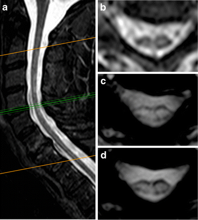
Axial 3D gradient-echo imaging for improved multiple sclerosis lesion  detection in the cervical spinal cord at 3T | SpringerLink
Axial 3D gradient-echo imaging for improved multiple sclerosis lesion detection in the cervical spinal cord at 3T | SpringerLink

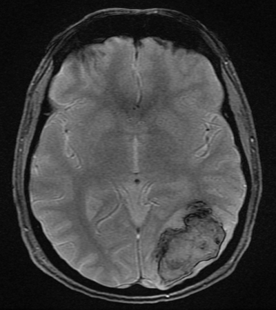
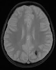
Axial T2* gradient echo-weighted brain magnetic resonance imaging of... |  Download Scientific Diagram
Axial T2* gradient echo-weighted brain magnetic resonance imaging of... | Download Scientific Diagram

Spoiled GRE - Questions and Answers ​in MRI

Axial gradient-echo MRI revealed that the right caudate nuclei and the... |  Download Scientific Diagram
Axial gradient-echo MRI revealed that the right caudate nuclei and the... | Download Scientific Diagram

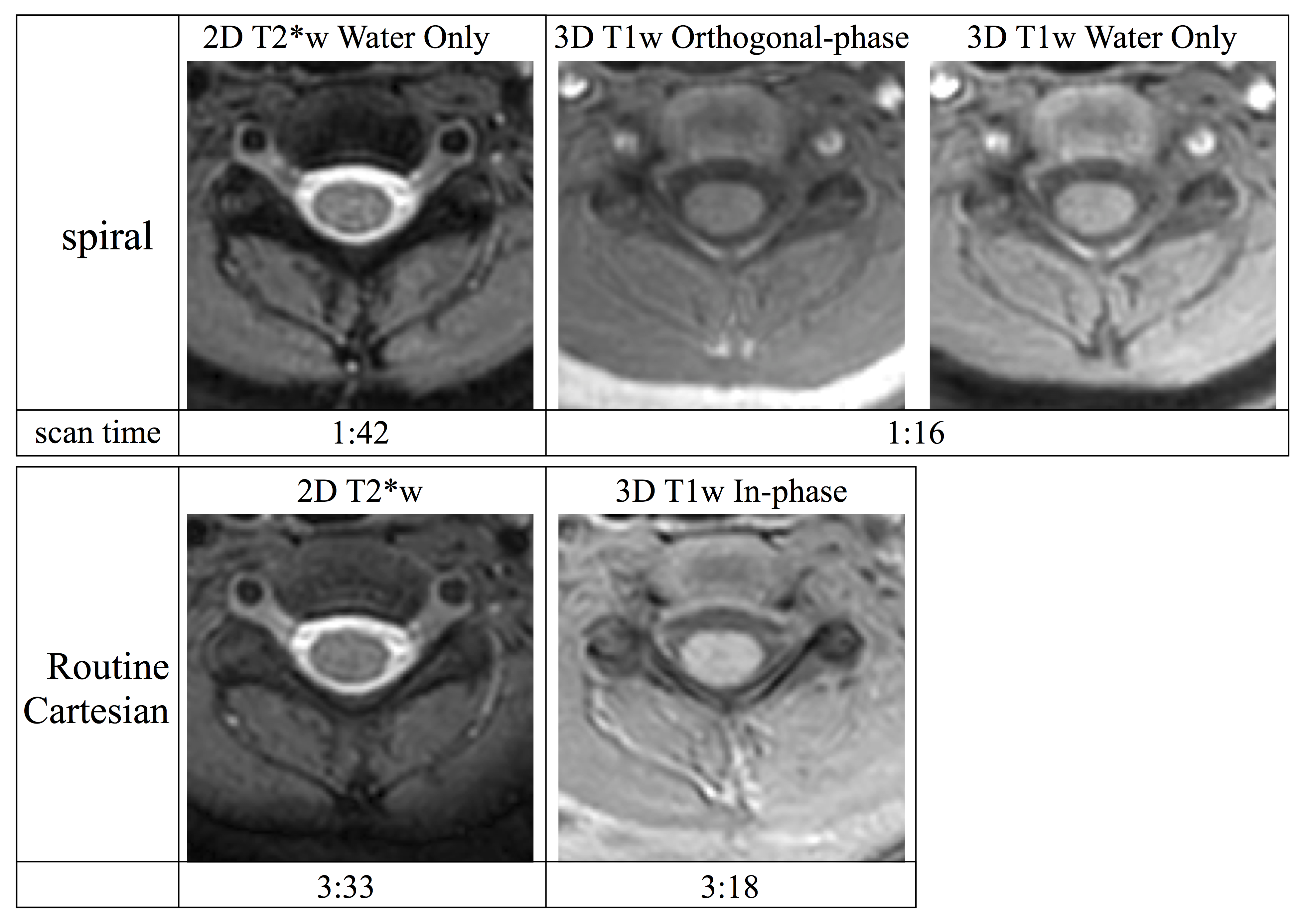
Axial Gradient Echo Spiral MRI of the Pediatric Spine

MR susceptibility contrast imaging using a 2D simultaneous multi-slice  gradient-echo sequence at 7T | PLOS ONE
MR susceptibility contrast imaging using a 2D simultaneous multi-slice gradient-echo sequence at 7T | PLOS ONE
Gradient echo imaging - Markl - 2012 - Journal of Magnetic Resonance  Imaging - Wiley Online Library
Gradient echo imaging - Markl - 2012 - Journal of Magnetic Resonance Imaging - Wiley Online Library

Gradient echo sequences | Radiology Reference Article | Radiopaedia.org

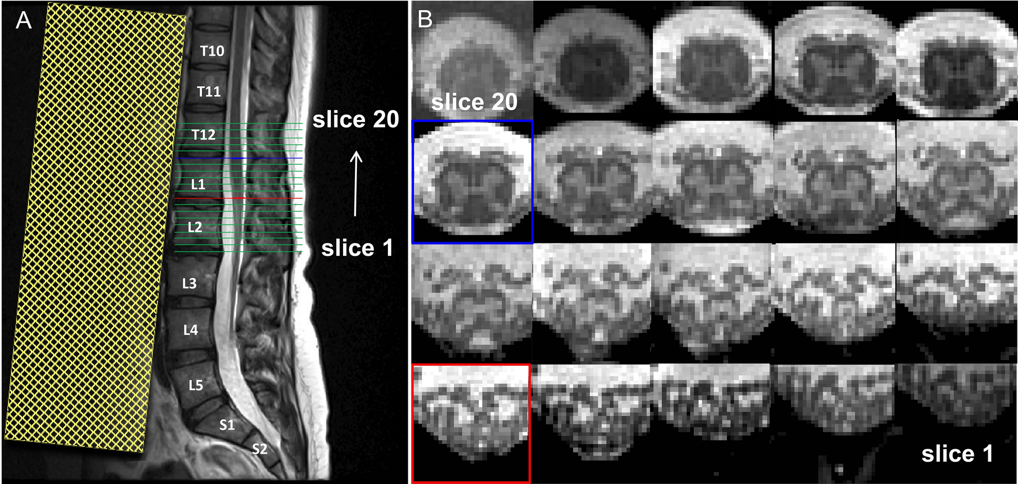
Optimized multi-echo gradient-echo magnetic resonance imaging for gray and  white matter segmentation in the lumbosacral cord at 3 T | Scientific  Reports
Optimized multi-echo gradient-echo magnetic resonance imaging for gray and white matter segmentation in the lumbosacral cord at 3 T | Scientific Reports

A–D) Gradient echo T2*-weighted axial MRI of the brain shows a rim of  hypointensity (consistent with the presence of haemosiderin deposits in the  leptomeninges. - ppt download
A–D) Gradient echo T2*-weighted axial MRI of the brain shows a rim of hypointensity (consistent with the presence of haemosiderin deposits in the leptomeninges. - ppt download

Comparison of axial gradient echo (GRE) images at 3 T (a) and 7 T (b)... |  Download Scientific Diagram
Comparison of axial gradient echo (GRE) images at 3 T (a) and 7 T (b)... | Download Scientific Diagram

Gradient Echo: Part I (Basic Principles) | Radiology Key

Comparison of MERGE and axial T2-weighted fast spin-echo sequences for  detection of multiple sclerosis lesions in the cervical spinal cord. |  Semantic Scholar
Comparison of MERGE and axial T2-weighted fast spin-echo sequences for detection of multiple sclerosis lesions in the cervical spinal cord. | Semantic Scholar